Example of a preliminary selection of images and reproductions of documents to be included in the publication:
Detail of the Photograph n.5 of David‘s technical examination report by the Scientific Police of Regional Unit. Document classified at the Superior Court of Florence, GIP section, file 12190/91
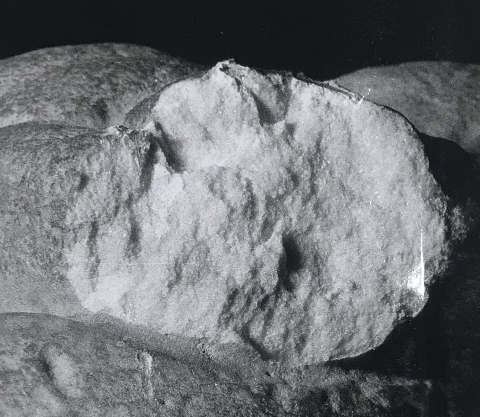
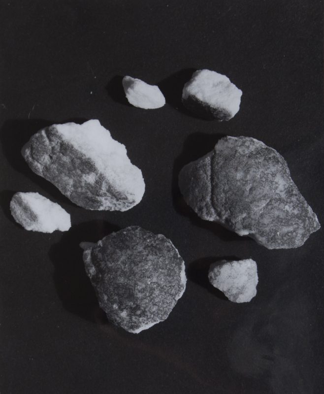
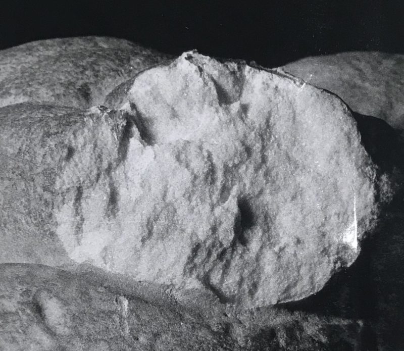
Detail of the photograph of seven fragments detached from the David’s toe. Document at the Photographic Archive of the Opificio delle Pietre Dure i Laboratori di Restauro. File n. 10579

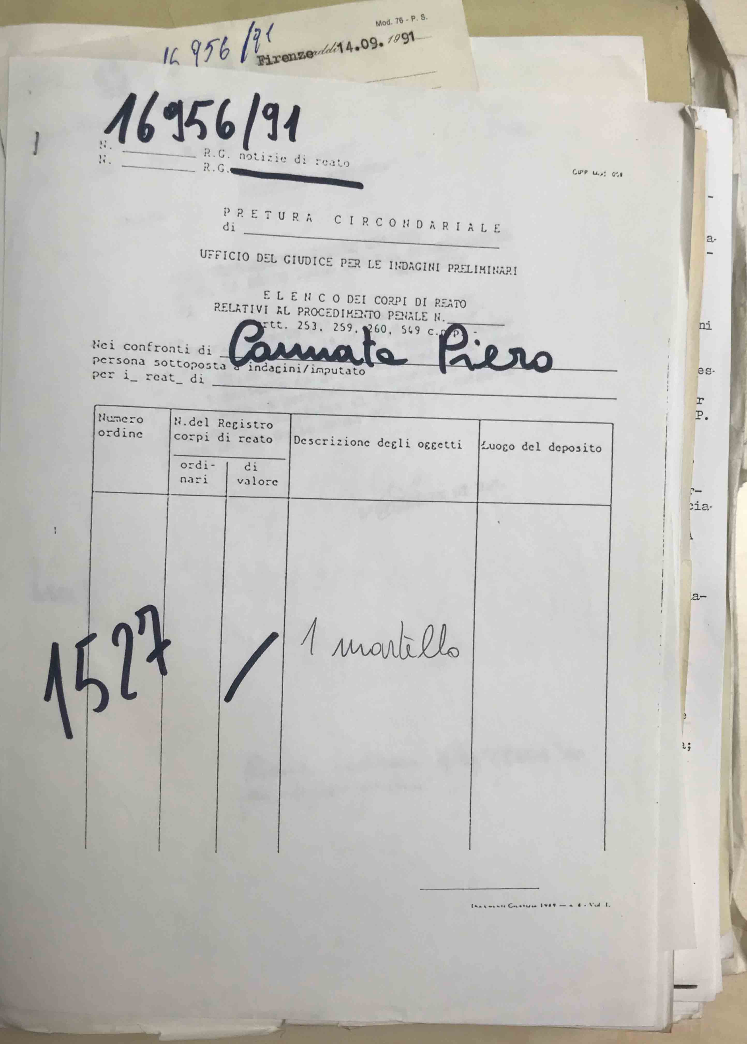
Report of seizure of a wooden hammer, owned by Piero Cannata. Florence Police Station. Document classified at the Superior Court of Florence, GIP section, file 12190/91
Introductory page of David’s technical examination report. Scientific Police of Regional Unit. Document classified at the Superior Court of Florence, GIP section, file 12190/91
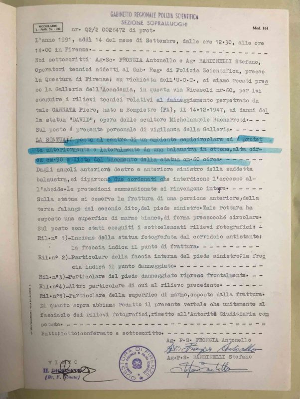
Written account of David’s technical examination report. Scientific Police of Regional Unit. Document classified at the Superior Court of Florence, GIP section, file 12190/91
Page number 3 of the Controlli analitici e chimico-fisichi sui frammenti di marmo proveniente dal dito del piede sinistro coordinated by Donato Attanasio (1991). Detail of the David’s sample used for measuruing the petographic properties (F17G, 20mg, 3 x 4 mm). Document provided by Susanna Bracci, scientific restorer of the Consiglio Nazionale delle Ricerche. The main information of the study was subsequently collected in AAVV. Exploring David. Diagnostic Tests and State of Conservation. Prato: Giunti Editore, 2004
Page number 9 of the Controlli analitici e chimico-fisichi sui frammenti di marmo proveniente dal dito del piede sinistro coordinated by Donato Attanasio (1991). Detail of the comparative graphs resulting of the analytical and chemical study. Document provided by Susanna Bracci, scientific restorer of the Consiglio Nazionale delle Ricerche. The main information of the study was subsequently collected in AAVV. Exploring David. Diagnostic Tests and State of Conservation. Prato: Giunti Editore, 2004

Photograph of some taròli. Photograph taken from the book AAVV. Exploring David. Diagnostic Tests and State of Conservation. Prato: Giunti Editore, 2004. Page 136

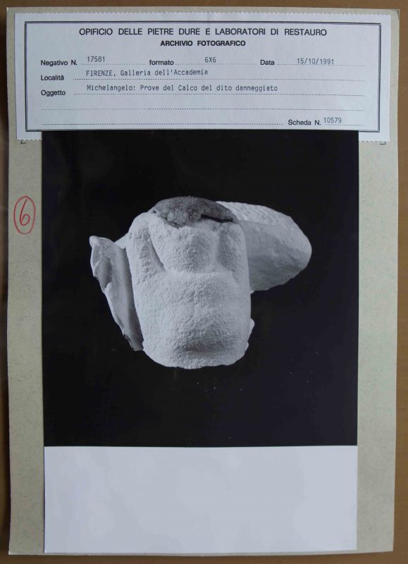
Tests for the reconstruction of David’s toe. Document at the Photographic Archive of the Opificio delle Pietre Dure i Laboratori di Restauro. File n. 10579

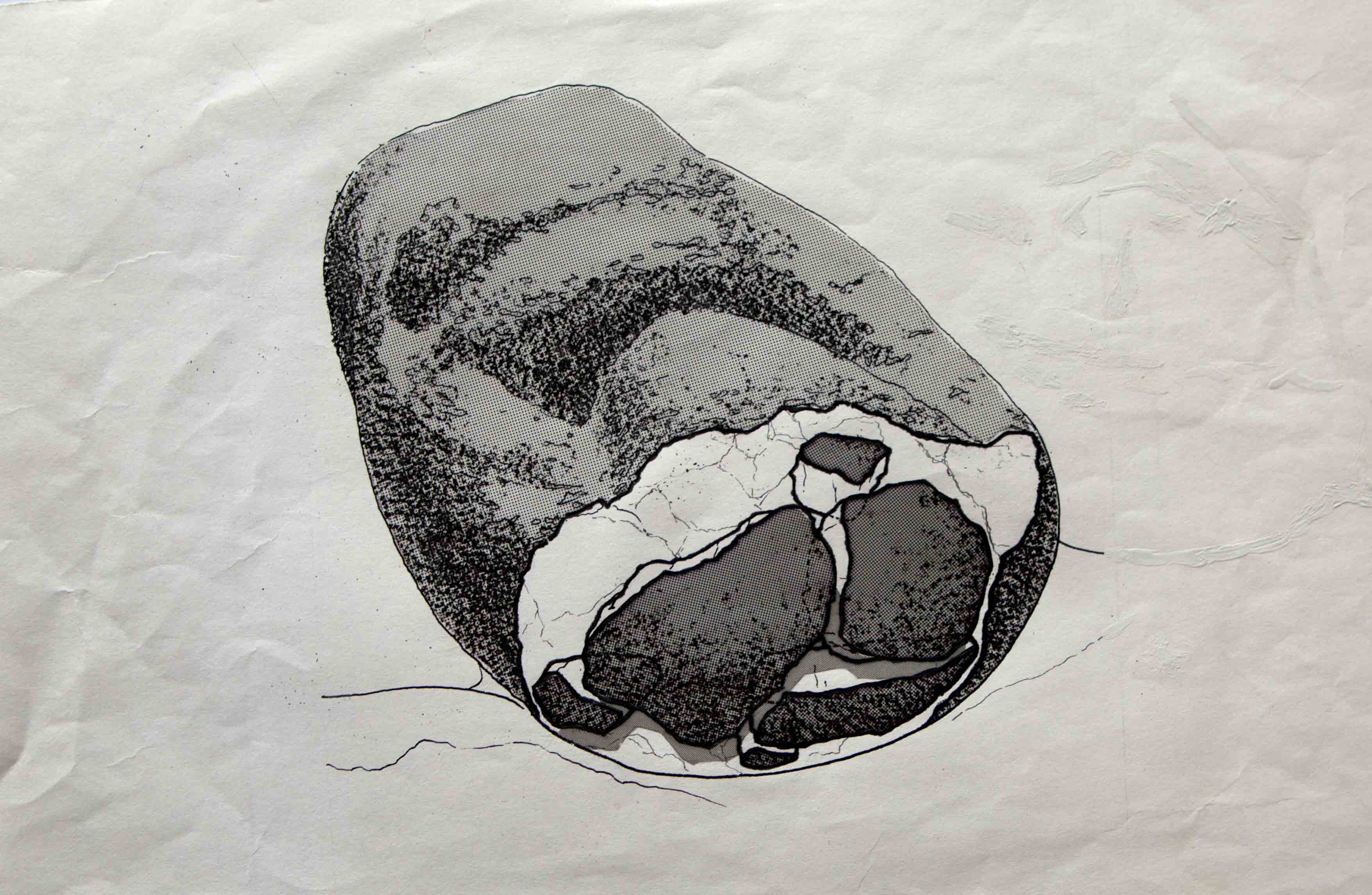
Visualization of the fragments’ placement. Document published at the article GIUSTI, Annamaria and NESTI, Roberto “‘David’ di Michelangelo: Un restauro in punta di piedi” in OPD. Rivista dell’Opificio delle Pietre Dure i Laboratori di Restauro. Orignal at the Photographic Archive of the Opificio delle Pietre Dure i Laboratori di Restauro. File n. 10579
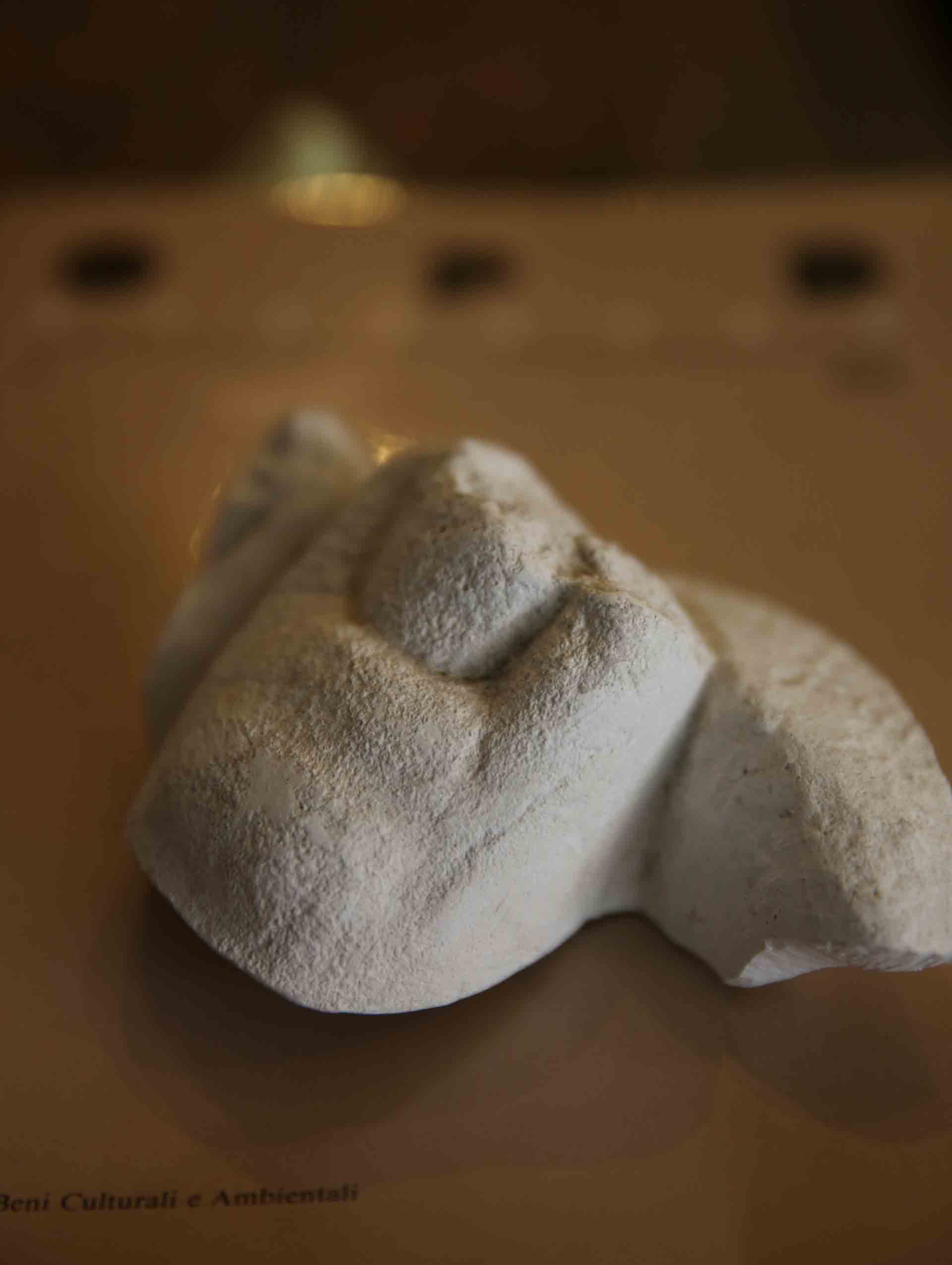
Plaster reproduction of the fractured toe. Photograph taken during a visit at the studio of the restorer Cinzia Parnigoni in July 2018

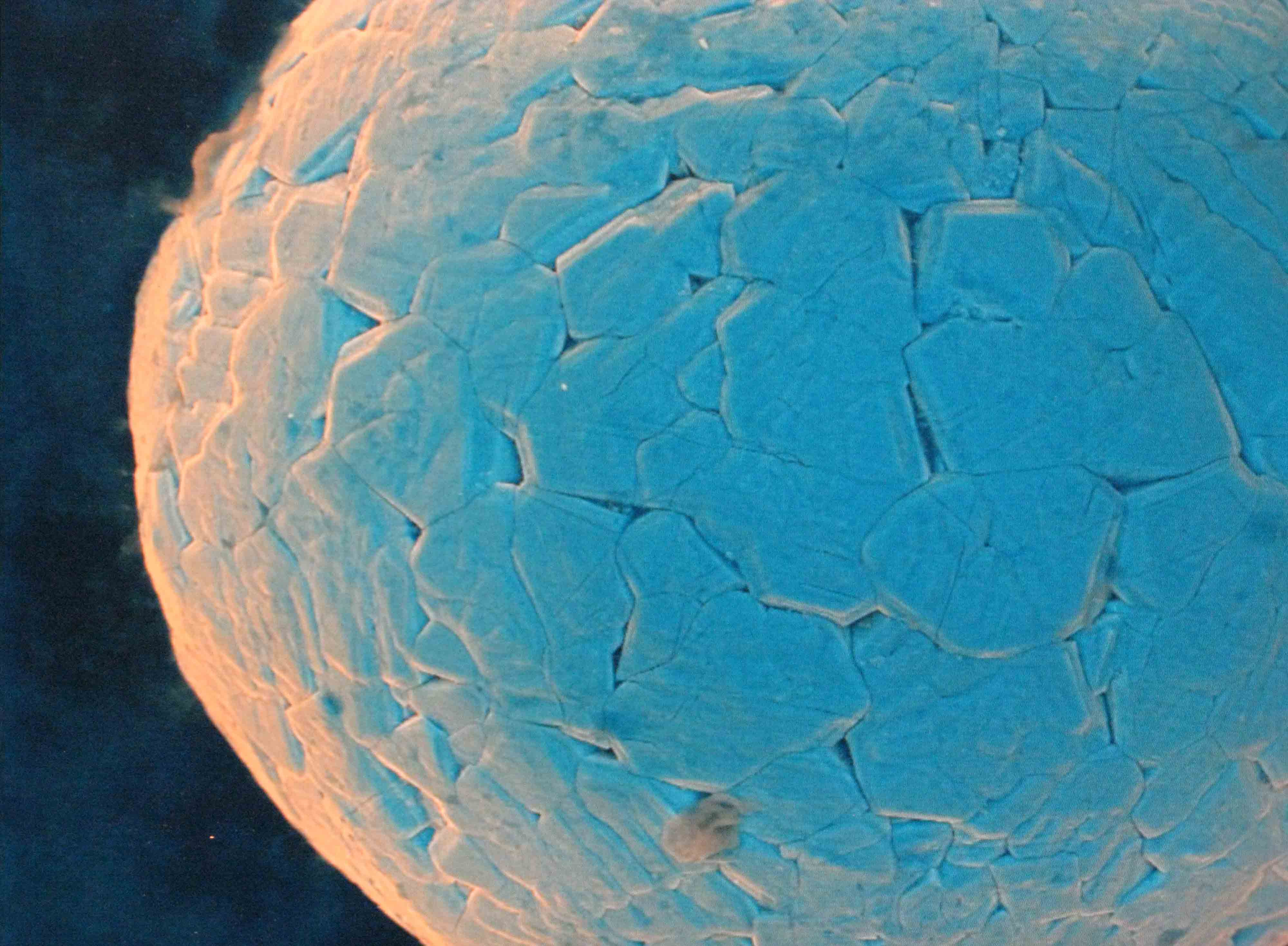
Thin section of the statue’s left toe showing intercrystalline porosity due to thermal dilation phenomena of the calcite crystals (6 X, crossed nicols). From Le caratteristiche mineralogiche, petrografiche e fisiche del marmo by Fabio Fratini. Later published at AAVV. Exploring David. Diagnostic Tests and State of Conservation. Prato: Giunti Editore, 2004
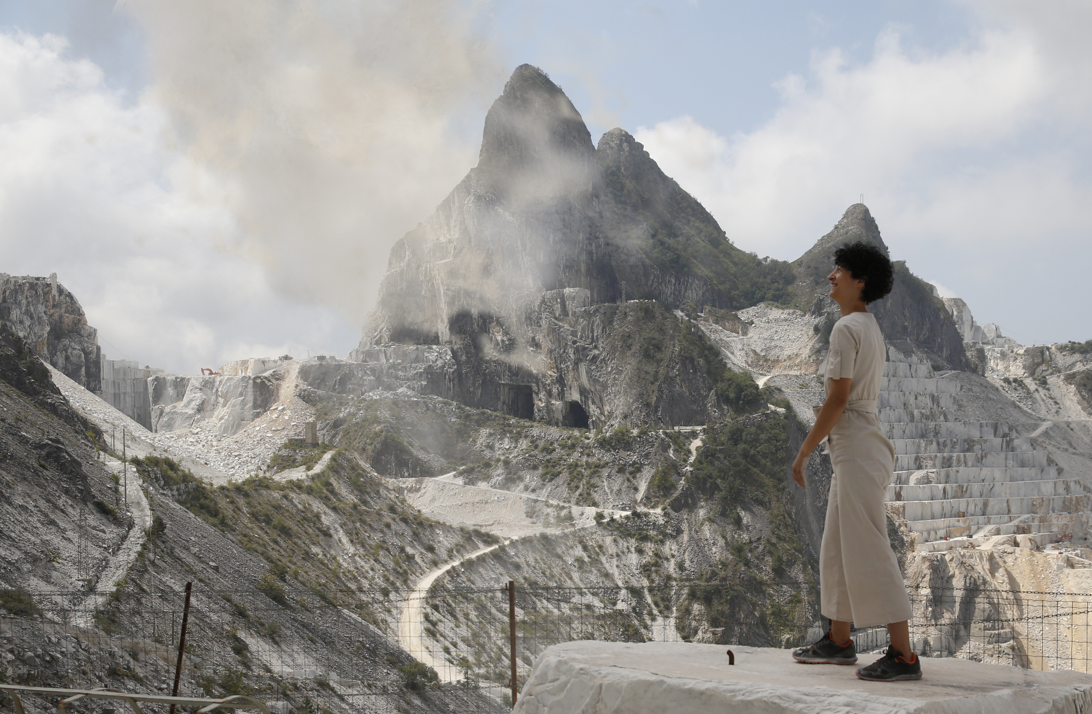
Throwing marble dust to form a cloud. Carrara, Michelangelo quarry, July 2018